OPIS PRZYPADKU
Refractory Central Serous Retinal Detachment in the Presence of Optic Disc Pit – Case Report
1
Department of Ophthalmology, Medical University of Wroclaw, Poland
Head: Professor Marta Misiuk-Hojło, MD, PhD
2
Ophthalmology Clinical Centre SPEKTRUM, Wroclaw, Poland
Data nadesłania: 04-03-2024
Data akceptacji: 02-04-2024
Data publikacji: 02-05-2024
Ophthalmology 2024;27(1):27-28
SŁOWA KLUCZOWE
STRESZCZENIE
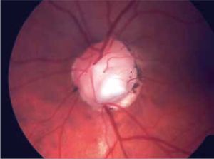
Among congenital optic disc anomalies are megalopapilla, optic nerve aplasia and hypoplasia (de Morsier’s syndrome) and optic disc excavations. The latter are attributed to abnormal fetal fissure’s closure. There is a wide spectrum of these anomalies ranging from single optic disc pit, to multiple pits, to morning glory syndrome. Excavated defects of the optic disc are usually associated with retinal detachment in the macular region. It is usually confined to the macular region though a subtotal or total retinal detachment may occur. It requires treatment (a surgical approach in most cases) though a spontaneous resolution of the subretinal fluid within a short period of the detachment onset has been observed. We report a case of unilateral central serous retinal detachment in the presence of a bilateral optic disc pit refractory to primary treatment.
REFERENCJE (10)
1.
Grear JN Jr.: Pits, or crater-like holes in the optic disc. Ach Ophthalmology. 1942; 28: 467–483.
2.
Dithmar S, Schuett F, Voelcker HE, et al.: Delayed sequential occurrence of perfluorodecalin and silicone oil in the subretinal space following retinal detachment surgery in the presence of an optic disc pit. Arch Ophthalmol. 2004 Mar; 122(3): 409–411.
3.
Becht C, Senn P, Lange AP: Delayed occurrence of subretinal silicone oil after retinal detachment surgery in an optic disc pit – a case report. Klin Monbl Augenheilkd. 2009 Apr; 226(4): 357–358. doi: 10.1055/ s-0028-1109249. Epub 2009 Apr 21.
4.
Brow GC, Shields JA, Goldberg RE: Congenital pits of the optic nerve head. II. Clinical studies in humans Ophthalmology 1980; 87: 51–65.
5.
Aobol WM, Boldi CF, Folk JC, et al.: Long term visual outcome in patients with optic nerve pit and serous retinal detachment of the macula. Ophthalmology. 1990; 79: 1539–1542.
6.
Gass JDM: Serous detachment of the macula secondary to congenital pit of the optic nerve head. Am J Ophthalmol. 1969; 67: 821–841.
7.
Lincoff H, Schiff W, Krivoy D, et al.: Optic coherenhce tomography of optic disc pit maculopatphy. Am J Ophthalmol. 1996; 122: 264–266.
8.
Akiyama H, Shimoda Y, Fukuchi M: Intravitreal gas injection without vitrectomy for macular detachment associated with an optic disc pit. Retina. 2013; 0: 1–6.
9.
Lincoff H, Kreissig I: Optical coherence tomography of pneumatic displacement of optic disc pit maculopathy. Br J Ophthalmol. 1998; 82: 367–372.
10.
Carlos AMN, Carlos AMJ: Vitrectomy and gas-fluid exchange for the treatment of serous macular detachment due to optic disc pit: long term evaluation. Arq Bras Ophthalmol. (Sao Paulo) 2013 May/June; Vol. 76: no. 3.
Przetwarzamy dane osobowe zbierane podczas odwiedzania serwisu. Realizacja funkcji pozyskiwania informacji o użytkownikach i ich zachowaniu odbywa się poprzez dobrowolnie wprowadzone w formularzach informacje oraz zapisywanie w urządzeniach końcowych plików cookies (tzw. ciasteczka). Dane, w tym pliki cookies, wykorzystywane są w celu realizacji usług, zapewnienia wygodnego korzystania ze strony oraz w celu monitorowania ruchu zgodnie z Polityką prywatności. Dane są także zbierane i przetwarzane przez narzędzie Google Analytics (więcej).
Możesz zmienić ustawienia cookies w swojej przeglądarce. Ograniczenie stosowania plików cookies w konfiguracji przeglądarki może wpłynąć na niektóre funkcjonalności dostępne na stronie.
Możesz zmienić ustawienia cookies w swojej przeglądarce. Ograniczenie stosowania plików cookies w konfiguracji przeglądarki może wpłynąć na niektóre funkcjonalności dostępne na stronie.


