Current issue
Archive
Online first
About the Journal
Editorial Board
Scientific Board
Reviewers
Abstracting and indexing
Publisher
Contact
Subscription
Ethical standards and procedures
Abstracting and indexing
For authors
Ethical standards and procedures
Publication charge
Books and Events
Books
Events
RESEARCH PAPER
Pre-Descemet Corneal Dystrophy – Changes in Corneal Morphology in Confocal Microscopy
1
Laser Eye Microsurgery Centre in Warsaw
2
Department of Ophthalmology Medical University of Warsaw
Publication date: 2024-08-19
Ophthalmology 2024;27(2):5-10

KEYWORDS
ABSTRACT
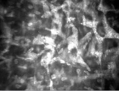
Purpose: To identify and describe the in-vivo microstructural changes in the cornea of patients with pre-Descemet’s membrane corneal dystrophy. Methods: Ten patients (20 eyes) were examined in the Laser Microsurgery Centre in Warsaw. The ophthalmic examination, including visual acuity and slit lamp biomicroscopy, was carried out. Because of suspicion of corneal dystrophy, in vivo confocal microscopy of the cornea was also performed. Results: Biomicroscopy revealed bilateral, dust-like fine opacities in the deep parts of the corneal stroma. Confocal microscopy showed pleomorphic structures (enlarged keratocytes) containing dense, hyperreflective, granular inclusions in the posterior stroma, next to the Descemet’s membrane. In 3 patients (6 eyes) punctate particles were seen extracellularly also in the mid stroma. Superficial and basal epithelial layers, anterior stroma and endothelium cells appeared normal. Conclusions: 1. In vivo confocal microscopy findings of cornea with pre-Descemet’s membrane dystrophy are characteristic. 2. In vivo confocal microscopy is a useful tool in diagnosing rare corneal dystrophies in cases where classical examination methods give inconclusive results.
REFERENCES (17)
1.
Afshari NA, Bouchard CS, Colby KA, et al.: Corneal dystrophies and ectasias. In: Weisenthal RW, ed. 2014–2015 Basic and Clinical Science Course, Section 8: External Disease and Cornea. San Francisco; American Academy of Ophthalmology. 2014: 253–287.
2.
Groenouw A: Knoetchenfoermige Hornhauttruebungen (Noduli corneae). Arch Augenheilkd. 1890; 21: 281–289.
3.
Weiss JS, Moller HU, Aldave AJ, et al.: IC3D classification of corneal dystrophies–edition 2. Cornea. 2015; 34: 117–159.
4.
Szaflik J, Szaflik JP, Słomińska M: Przyżyciowa ocena struktury rogówki po zabiegach keraptoplastyki drążącej w mikroskopii konfokalnej, ze szczególnym uwzględnieniem aspektu wtórnego unerwienia przeszczepionej rogówki – doniesienia wstępne. [In vivo confocal microscopy of corneal grafts after penetratingkeratoplasty, with particular accent on graft reinnervation – initial report]. Okulistyka. 2003; 2: 32–39.
5.
Maeder G, Danis P: On a new form of corneal dystrophy (deep filiform dystrophy of the cornea) associated with keratoconus. Ophthalmologica. 1947; 114: 246.
6.
Franceschetti A, Maeder G: Deep corneal dystrophy in a case of congenital ichthyosis. Bull Soc Fr Ophtalmol. 1954; 67: 146.
7.
Collier M: Hereditary nature of the punctate pre-descemet dystrophy. Bull Soc Fr Ophtalmol. 1964; 64: 731.
9.
Alio del Bario JL, Chung DD, Al-Shymali O, et al.: Punctiform and Polychromatic Pre-Descemet Corneal Dystrophy: Clinical Evaluation and Identification of the Genetic Basis. Am J Ophthalmol. 2020 April; 212: 88–97.
10.
Curran RE, Kenyon KR, Green WR: Pre-Descemet’s membrane corneal dystrophy. Am J Ophthalmol. 1974; 77: 711–716.
11.
Kempster RC, Hirst LW, de la Cruz Z, et al.: Clinicopathologic study of the cornea in X-linked ichthyosis. Arch Ophthalmol. 1997; 115: 409–415.
12.
Shi H, Qi XF, Liu TT, et al.: In vivo confocal microscopy of pre-Descemet corneal dystrophy associated with X-linked ichthyosis: a case report. BMC Ophthalmol. 2017 Mar 16; 17(1): 29.
13.
Malhotra C, Jain AK, Dwivedi S, et al.: Characteristics of Pre-Descemet Membrane Corneal Dystrophy by Three Different Imaging Modalities-In Vivo Confocal Microscopy, Anterior Segment Optical Coherence Tomography, and Scheimpflug Corneal Densitometry Analysis. Cornea. 2015; 34: 829–832.
14.
Alafaleq M, Georgeon C, Grieve K, et al.: Multimodal imaging of pre-Descemet corneal dystrophy. Eur J Ophthalmol. 2020 Sep; 30(5): 908–916.
15.
Sonkeshri A, Agrawal D, Chatterjee S, et al.: Pre-Descemet’s corneal dystrophy. Indian Journal of Ophthalmology - Case Reports. 2021 Jan–Mar; 1: 8.
16.
Yeh SI, Liu TS, Ho CC, et al.: In vivo confocal microscopy of combined pre-descemet membrane corneal dystrophy and fuchs endothelial dystrophy. Cornea. 2011 Feb; 30(2): 222–224.
17.
Kontadakis GA, Kymionis GD, Kankariya VP, et al.: Corneal confocal microscopy findings in sporadic cases of pre-descemet corneal dystrophy. Eye Contact Lens. 2014 Mar; 40(2): e8–e122017 Mar 16; 17(1): 29.
| eISSN: | 1689-362X |
| ISSN: | 1505-2753 |
We process personal data collected when visiting the website. The function of obtaining information about users and their behavior is carried out by voluntarily entered information in forms and saving cookies in end devices. Data, including cookies, are used to provide services, improve the user experience and to analyze the traffic in accordance with the Privacy policy. Data are also collected and processed by Google Analytics tool (more).
You can change cookies settings in your browser. Restricted use of cookies in the browser configuration may affect some functionalities of the website.
You can change cookies settings in your browser. Restricted use of cookies in the browser configuration may affect some functionalities of the website.